|
|
Localized Fibrous Tumor of the Pleura
Solitary Fibrous Tumor of the Pleura, Benign Mesothelioma, Pleural Fibroma
General Considerations
- Rare, mesenchymal primary tumors of the visceral pleura less common than diffuse malignant mesothelioma
- Localized form is less common and not related to asbestos exposure or smoking
- LFTP can be either benign or malignant but is much more often benign (7:1)
- Usually patients are >50 years old
- Most occur in the inferior portions of the hemithoraces
Clinical Findings
- May be asymptomatic and found incidentally
- May produce chest wall pain, cough, hemoptysis, hypertrophic osteoarthropathy or hypoglycemia be cause they secrete insulin-like proteins
Imaging Findings
- Conventional radiography
- Well-circumscribed soft tissue mass with sharp margin applied to a pleural surface, including the fissures
- Angle it makes with chest wall may be acute or obtuse
- They can grow very large and occupy half the hemithorax
- CT
- Soft-tissue mass applied to pleural surface
- May have a lobulated contour
- Avidly enhances but may have lower attenuation areas of necrosis or hemorrhage, especially in larger lesions
- At least one angle with pleura is more often acute
- May have small effusion
- Calcification is rare
- May have a pedicle that attached tumor to pleura
- Malignant lesions are more often large and have necrosis
- MRI
- T1 low to intermediate and T2 low
- Intense enhancement with Gadolinium
Differential Diagnosis
- Biopsy is required to differentiate benign from malignant form
Treatment
- Resectable for cure
- If totally excised, they usually do not recur
Complications
- Hypertrophic osteoarthropathy
- Hypoglycemia
- May rarely undergo malignant transformation or recur locally
Prognosis
- 12% recurrence rate has been cited with an 88% cure overall with resection


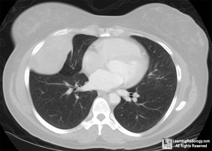
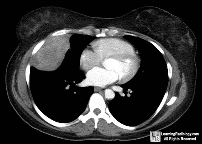
Localized Fibrous Tumor of the Pleura. Upper photo. Large, pleural-based soft tissue mass abuts
pleura in right lower lung with an acute angle (white arrow) and obtuse angle (yellow arrow) where it meets
the chest wall. Lower photo: Mediastinal windows show heterogeneous nature of the contrast-enhancing mass
with some areas of lower attenuation (blue arrows) most likely representing necrosis.
For these same photos without the arrows, click here and here
For more information, click on the link if you see this icon 
eMedicine. Imaging of Localized Fibrous Tumor of the Pleura. MA Meziane and O Lababede
Localized Fibrous Tumors of the Pleura. ML. Rosado-de-Christenson, GF Abbott, HP McAdams, TJ Franks, and JR Galvin. May 2003 RadioGraphics, 23, 759-783
|
|
|
